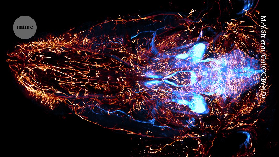
A new imaging technique successfully maps the nerve connections from a mouse's brain and spinal cord to the body with micrometre-scale resolution. Previous attempts focused on the brain's connectome, but mapping peripheral nerves posed challenges. Researchers use a custom-built microscope to scan tissue after treating it with chemicals that remove obstructive components. This method captures detailed 3D images by pushing the mouse towards a slicing blade. The speed of this method reduces the likelihood of mechanical issues, making it a significant advancement in connectomics beyond the brain.
"A speedy imaging method can map the nerves running from a mouse's brain and spinal cord to the rest of its body at micrometre-scale resolution, revealing details such as individual fibres travelling from a key nerve to distant organs."
"To prepare a mouse's body for the scan, researchers treat it with chemicals that make its tissues transparent by removing fat, calcium and other components that block light."
"The authors say the method is much faster than other techniques, which require frequent pauses. A slower system would take months or years, increasing the chance of mechanical failure, signal loss or sample degradation."
Read at Nature
Unable to calculate read time
Collection
[
|
...
]