"Rice weevil (Sitophilus oryzae) on a grain of rice. Photographer Zhang You had shot Rice weevils before, but never one with its wings spread'. This one was naturally preserved on a windowsill, perhaps in a final attempt to escape.' Photograph: Zhang You/Nikon Small World Colonial algae (Volvox) spheres in a drop of water. Volvox is a genus of green algae, forming spherical colonies of up to 50,000 cells. Photograph: Dr Jan Rosenboom/Nikon Small World"
"Heart muscle cells with chromosomes condensed after cell division, using 100x objective lens magnification. Photograph: Dr James Hayes/Nikon Small World Spores (blue/purple structures) of a small tropical fern (Ceratopteris richardii), widely studied to understand plant evolution. Photograph: Dr Igor Siwanowicz/Nikon Small World Rat liver cells, beautifully depicted here, are used in research to understand the organ's metabolism and detoxification. Photograph: Dr Francisco Lazaro-Dieguez/Nikon Small World iPSC-derived sensory neurons, labelled to show tubulin and actin, under immunofluorescence microscopy. Photograph: Stella Whittaker/Nikon Small World"
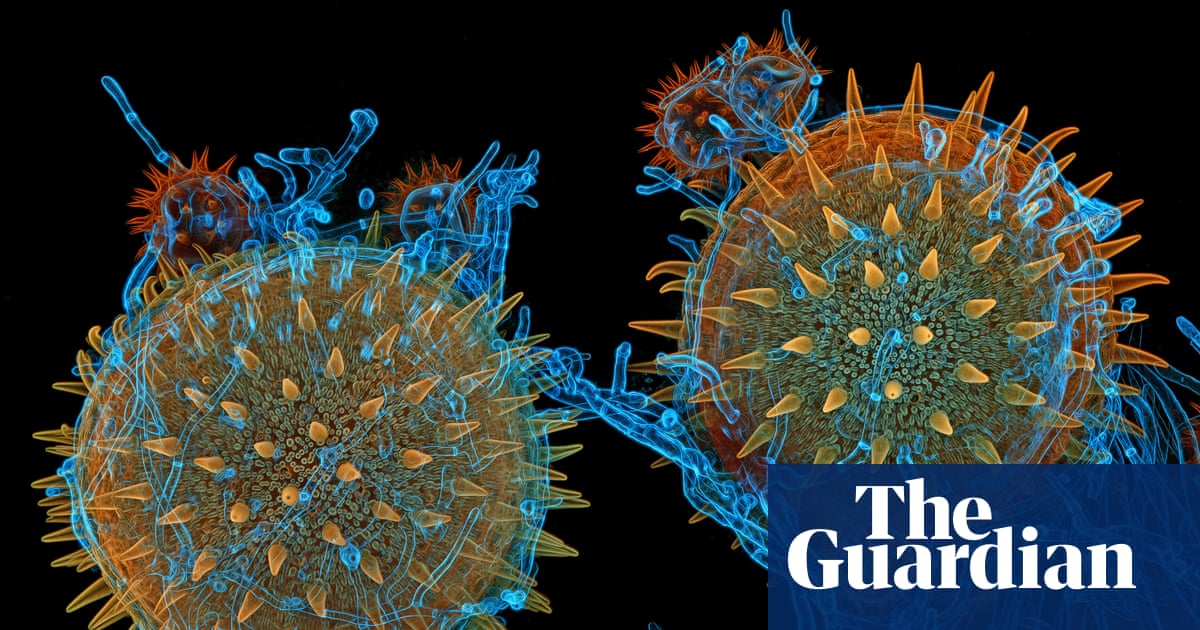
"Mallow pollen germinating on stigma, the process in flowering plants, while being parasitised by a filamentous fungus. Photograph: Dr Igor Siwanowicz/Nikon Small World A fungus Talaromyces purpureogenus known for its red, diffused pigment. Photograph: Wim van Egmond/Nikon Small World Heart muscle cells (iPSC-derived) showing condensed chromosomes in metaphase. Photograph: Dr Dylan Burnette & Dr James Hayes/Nikon Small World Sunflower trichomes are hair-like outgrowths on the plant's surface. Photograph: Marek Mis/Nikon Small World"
A collection of high-magnification images presents diverse microscopic subjects across biological kingdoms. Insect detail appears in a rice weevil captured on a grain with wings spread. Colonial green algae (Volvox) form spherical colonies visible in a drop of water. Plant structures include pollen caught in a spider web, mallow pollen germinating on a stigma, and sunflower trichomes. Fungi appear as pigmented Talaromyces and filamentous parasites on pollen. Cellular and tissue-level images show heart muscle cells during division, iPSC-derived neurons and sensory cells labelled for tubulin and actin, rat liver cells, and a cancer cell with highlighted actin cytoskeleton and endoplasmic reticulum.
Read at www.theguardian.com
Unable to calculate read time
Collection
[
|
...
]