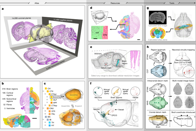
Brain stereotaxic atlases are critical for spatial localization and understanding brain structure organization. Recent advancements in spatial transcriptomics and neural circuit tracing have increased the need for precise cell localization within the brain. Traditional rodent brain atlases rely on manual annotations of Nissl-stained sections, which create gaps that hinder continuous anatomical observation and 3D reconstruction accuracy. To address these constraints, a common coordinate framework (CCF) has been developed using mouse brain tissue autofluorescence, though its reliance on low-resolution data raises concerns about accuracy in anatomical delineation and cellular positioning.
"Brain stereotaxic atlases are essential for determining spatial locations and understanding organizational principles in the brain, but traditional methods face significant limitations."
"Traditional rodent brain atlases use Nissl-stained sections that are manually annotated, leading to challenges in observing continuous anatomical changes along the axial direction."
"The construction of a common coordinate framework (CCF) aims to address limitations of 2D atlases by facilitating 3D brain mapping of neural circuits and multi-omics datasets."
"The CCF utilizes autofluorescence of the mouse brain tissue for anatomical delineations, but the dataset's lower resolution raises concerns over the accuracy of cellular localization."
#brain-mapping #spatial-transcriptomics #neuroanatomy #common-coordinate-framework #3d-reconstruction
Read at Nature
Unable to calculate read time
Collection
[
|
...
]