""It's exciting," says Dr. Stephen Waxman, a professor at Yale School of Medicine who was not involved in the research. Currently, prospective pain drugs are typically tested in animals whose responses are often different than a human's and in individual nerve cells, which may not reflect the behavior of entire brain networks. With this new system, known as a brain assembloid, "we have a miniature nervous system that might be a very useful platform," Waxman says."
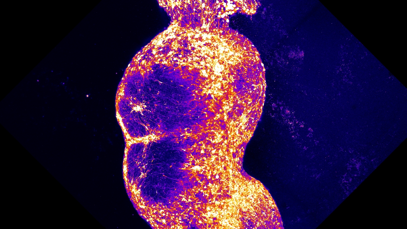
"To approximate this pathway in the lab, Pasca's team created four different brain organoids, spherical clumps of human nerve cells that grow in a dish. The team coaxed each organoid to resemble one specific type of brain or spinal tissue found along the pain pathway. "And then we put them together, really put them in close proximity, and watched them as they connected with each other," Pasca says."
"After more than six months developing in the lab, the resulting assembloid had created a pathway linking the four organoids. The nerve cells also spontaneously began "working in a coordinated fashion across the four parts of this assembloid," Pasca says."
"A pathway with several stops, the model is the result of an effort to re-create the signaling chain that occurs after exposure to painful stimuli, says Dr. Sergiu Pasca, a professor at Stanford University who led the project."
A Stanford research team has successfully created a laboratory model mimicking the human pain pathway by developing four key clusters of human nerve cells in a dish, referred to as a brain assembloid. This innovation allows for a more accurate assessment of pain syndromes and potential analgesic drugs compared to traditional animal testing. The team developed distinct organoids representing various nervous system tissues and connected them, resulting in spontaneous coordination among the nerve cells. This miniature nervous system could serve as a more relevant platform for drug testing than individual cells or animal models.
Read at www.npr.org
Unable to calculate read time
Collection
[
|
...
]