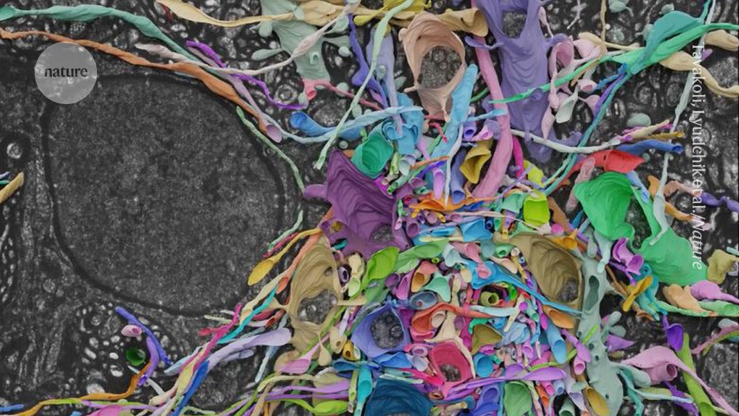
"Scientists have developed a method to visualize mouse brain cell patterns, inflating samples to 16 times their size for detailed mapping with standard light microscopes."
A novel method allows scientists to map intricate brain cell patterns in mouse tissue using a standard light microscope. By inflating samples 16 times their original size with special gels, researchers can visualize individual neuron connections and synapses more clearly. This method, which combines traditional microscopy with artificial intelligence for enhanced imaging, presents a faster, cost-effective alternative to electron microscopy. It could offer rich, colorful insights into brain connectivity that were previously unseen and marks a significant advancement in neurological studies.
Read at Nature
Unable to calculate read time
Collection
[
|
...
]