"In brief, circular (3-mm diameter) craniotomies were centred over ALM (2.5 mm anterior and 1.5 mm lateral from Bregma). We expressed the soma-targeted opsin ST-ChrimsonR in excitatory neurons by injecting a virus (10 12 titre; AAV2/2 camKII-KV2.1-ChrimsonR-FusionRed; Addgene, plasmid, catalogue no. 102771) into the craniotomy, 400 µm below the dura (five to ten sites, 20-30 nl each), centred in the craniotomy and spaced by approximately 500 μm between injection sites."
"The craniotomy was covered by a cranial window composed of three layers of circular glass (total thickness 450 μm). The diameter of the bottom two layers was 2.5 mm. The top layer was 3 mm or 3.5 mm and rested on the skull. The window was cemented using cyanoacrylate glue and dental acrylic (Lang Dental). A customized headbar was attached using cyanoacrylate glue and dental cement."
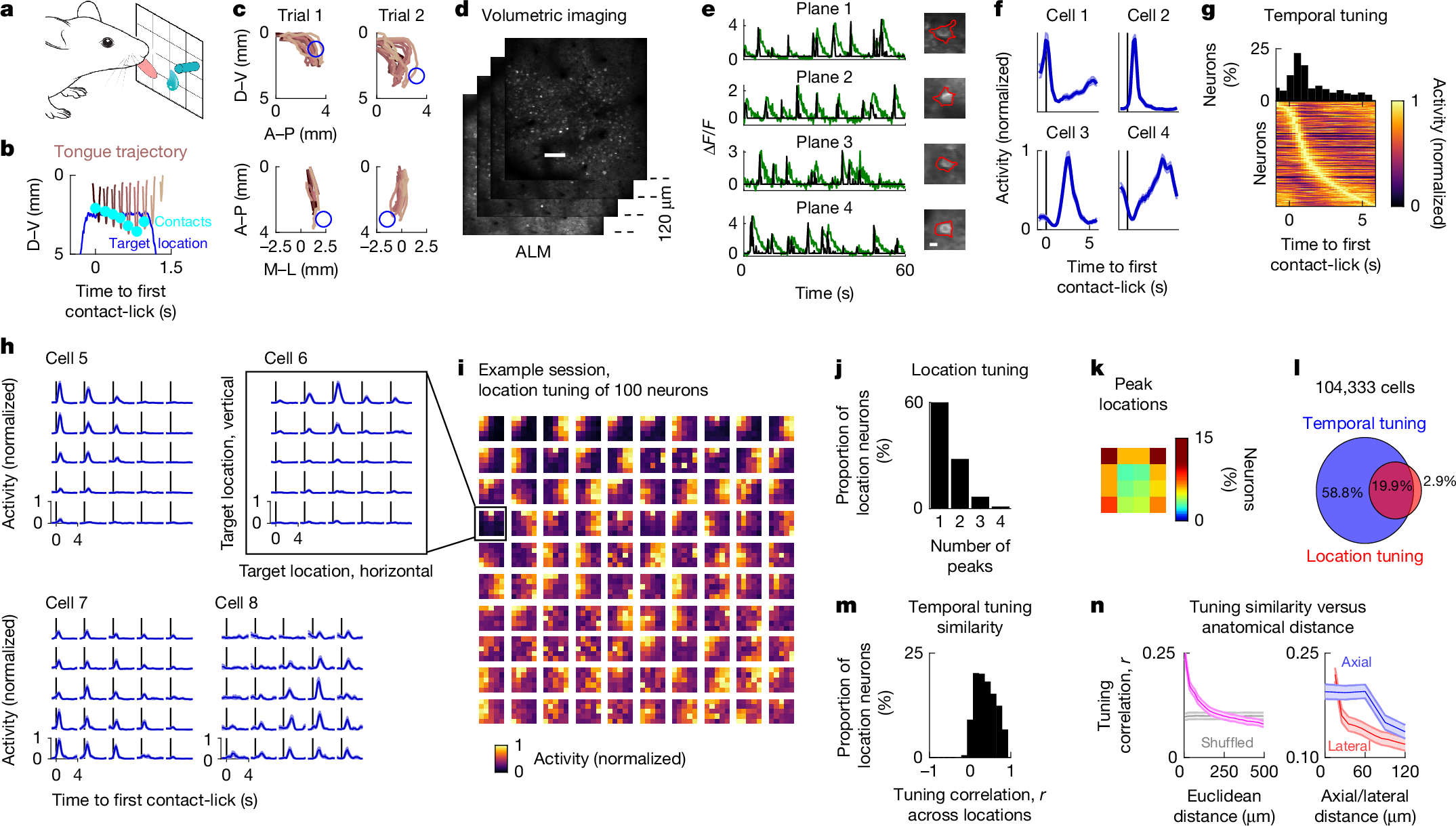
Nineteen mice (60–240 days old) of either sex were used, with fifteen for imaging and behavioral experiments and four for behavioral data only. The genotype was CamK2a-tTA × Ai94 (TITL-GCaMP6s) × slc17a7 IRES Cre, providing widespread GCaMP6s expression in excitatory cortical neurons. Mice were housed on a 12:12 reverse light:dark cycle with behavioral experiments during the dark phase and maintained at 21±1°C and 50±10% relative humidity. Circular 3-mm craniotomies were centered over ALM. ST-ChrimsonR was expressed via AAV injections 400 µm below the dura at multiple sites. Cranial windows comprised three glass layers (450 µm total) and a customized headbar was attached. Water restriction (1 ml/day) began 3–4 weeks after viral expression.
Read at Nature
Unable to calculate read time
Collection
[
|
...
]