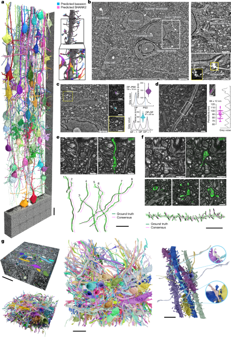
"Light microscopy holds considerable potential for unifying synapse-level circuit reconstruction with in-depth molecular characterization, despite limits in resolution that hinder identifying structures."
"Electron microscopy (EM) is crucial for dense connectomic analysis, enabling comprehensive reconstruction of cellular circuit components due to its nanometre-scale resolution."
The article discusses the intricate arrangement of neurons in the brain and the importance of imaging techniques to decode this organization. While light microscopy shows promise for circuit reconstruction, its resolving power is inadequate for densely packed cellular structures. Electron microscopy (EM) emerges as the preferred method due to its nanometre-scale precision, allowing for detailed mapping of neural connectivity. Recently, advancements in automated data collection and deep learning have enhanced EM's application in connectomics across various organisms, although it requires complementary light microscopy to relate structural and molecular information.
Read at Nature
Unable to calculate read time
Collection
[
|
...
]