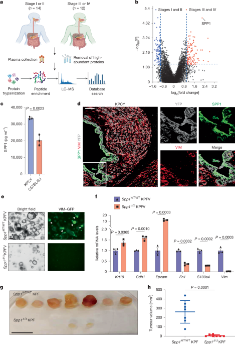
"PDAC is one of the leading causes of cancer-related mortality worldwide, with a 5-year relative survival rate of only 13%5. To identify factors that control the progression of pancreatic cancer, we compared protein levels in the plasma of patients with early-stage (stage I or II) or late-stage (stage III or IV) PDAC (Fig. 1a and Supplementary Table 1). The abundance of SPP1 was significantly increased at late stages (Fig. 1b)."
"d, Immunofluorescence analysis of VIM (red), YFP (grey) and SPP1 (green) in KPCY tumours. Scale bar, 50 μm. e, Bright-field and fluorescence images of VIM-GFP in Spp1WT/WT KPFV and Spp1Δ/Δ KPFV organoids. Arrowheads indicate VIM-GFP+ cells. Scale bar, 50 μm. f, RT -qPCR analysis of expression of the epithelial markers Krt19, Cdh1 and Epcam and the mesenchymal markers Fn1, S100a4 and Vim between Spp1WT/WT KPFV and Spp1Δ/Δ KPFV organoids ( n = 3 biological replicates), normalized using Spp1WT/WT KPFV values."
Pancreatic ductal adenocarcinoma (PDAC) exhibits a 5-year relative survival rate near 13%. Proteomic profiling compared plasma proteins from patients with early-stage (I–II) and late-stage (III–IV) PDAC to identify progression-associated factors. SPP1 abundance was significantly increased in late-stage patient plasma and was elevated in Pdx1-cre;KrasLSL-G12D/+;Trp53fl/fl;Rosa26LSL-YFP (KPCY) mice compared with wild-type controls. Immunofluorescence assessed VIM, YFP and SPP1 localization in KPCY tumours. Organoid experiments used VIM-GFP Spp1WT/WT and Spp1Δ/Δ KPFV lines and RT–qPCR to compare epithelial markers (Krt19, Cdh1, Epcam) and mesenchymal markers (Fn1, S100a4, Vim). Subcutaneous tumour formation and tumor sizes were evaluated between genotypes.
Read at Nature
Unable to calculate read time
Collection
[
|
...
]