An international team from University College London has published the Human Organ Atlas, a comprehensive 3D imaging platform of human organs created using HiPCT, an extremely powerful X-ray technique. The atlas provides cellular-level detail without damaging tissue, connecting macroscopic anatomy with microscopic histological information. This five-year project enables researchers to study complex diseases like hypertension, diabetes, cancer, and COVID-19 by examining how organ failures relate across multiple systems. The platform incorporates data from deceased donors and represents a significant advancement in biomedical research. Initial discoveries from COVID-19 patient data revealed previously unknown vascular lesions, demonstrating the atlas's research potential. Managing massive datasets, some exceeding terabytes, presented major technical challenges for the team of over 20 researchers.
"The project, a kind of Google Earth of the body, promises to revolutionize biomedical research and the study of diseases, such as hypertension, diabetes, cancer and COVID-19, by linking multisystem failures, anatomical variations and patterns associated with complex pathologies in organs of the same patient."
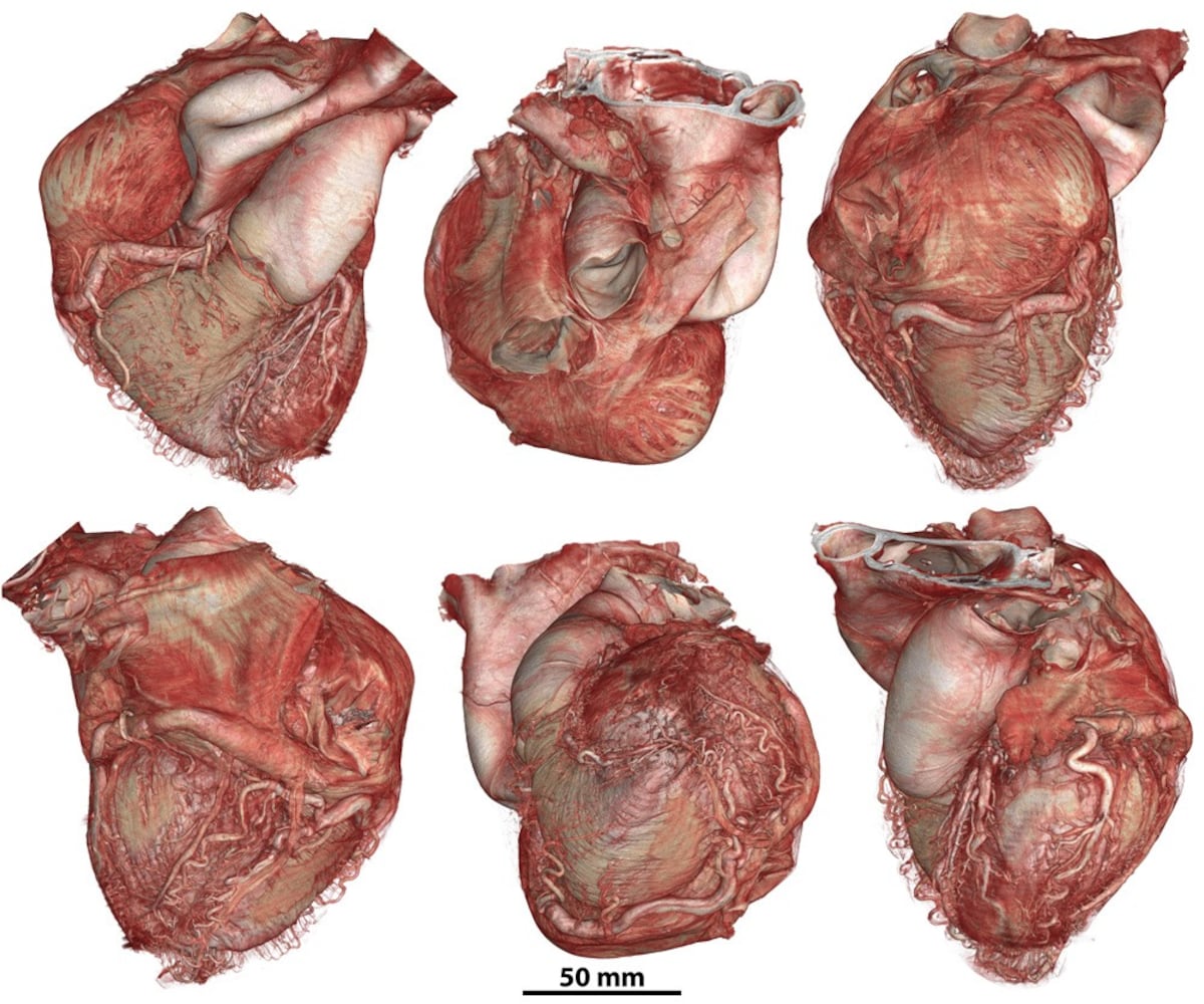
"This multiscale capability makes this atlas a unique resource, capable of connecting macroscopic anatomy with 3D histological details, say the researchers, who published their work in the journal Science Advances."
"The researcher recalls that some of the initial information shared in the atlas about patients who died from COVID-19 led to publications revealing previously unseen microscopic vascular lesions. That and other discoveries became the foundation for building something bigger."
Read at english.elpais.com
Unable to calculate read time
Collection
[
|
...
]